






New
Compare
Report Abuse
Yonker Premium Diagnostic UItrasound System Revo 9
Available in stock
Yonker Premium Diagnostic UItrasound System Revo 9 is a Yonker medical product for professional clinical, diagnostic, monitoring, imaging, treatment, or supervised healthcare use. Contact Scientific China for configuration, certification, MOQ, availability, and quotation.

Scientific China
StorePeople Also Viewed
-
 C3510 - Auto Coagulation Analyzer
-
 BeneFusion e Series - Infusion Pump
-
 3 Part Differential Hematology Analyzer - BC-20
-
 BeneFusion 5 Series - Infusion System
-
 A3 Anesthesia Workstation
-
 Infusion Pump Disposables
-
 Defibrillator Monitor - BeneHeart D30
-
 Crius V6 ICU Ventilator – Advanced Mechanical Ventilation with HFNC & Turbine Technology
Additional information
| Product Category | Ultrasound Systems |
|---|
Questions & Answers
Loading...
Product Enquiry
Product Enquiry
Description
Yonker Premium Diagnostic UItrasound System Revo 9
Product overview
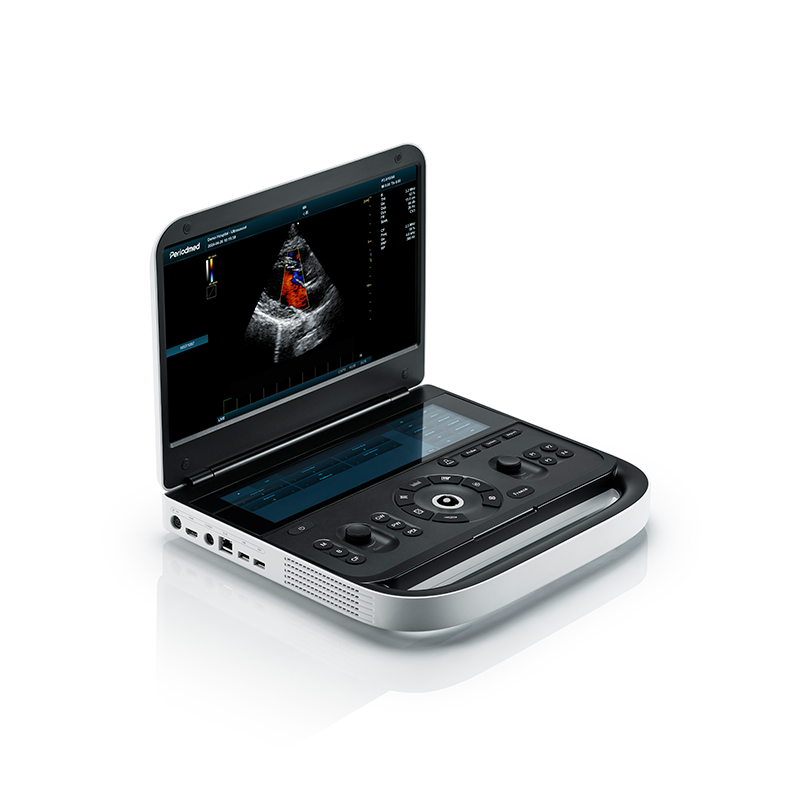
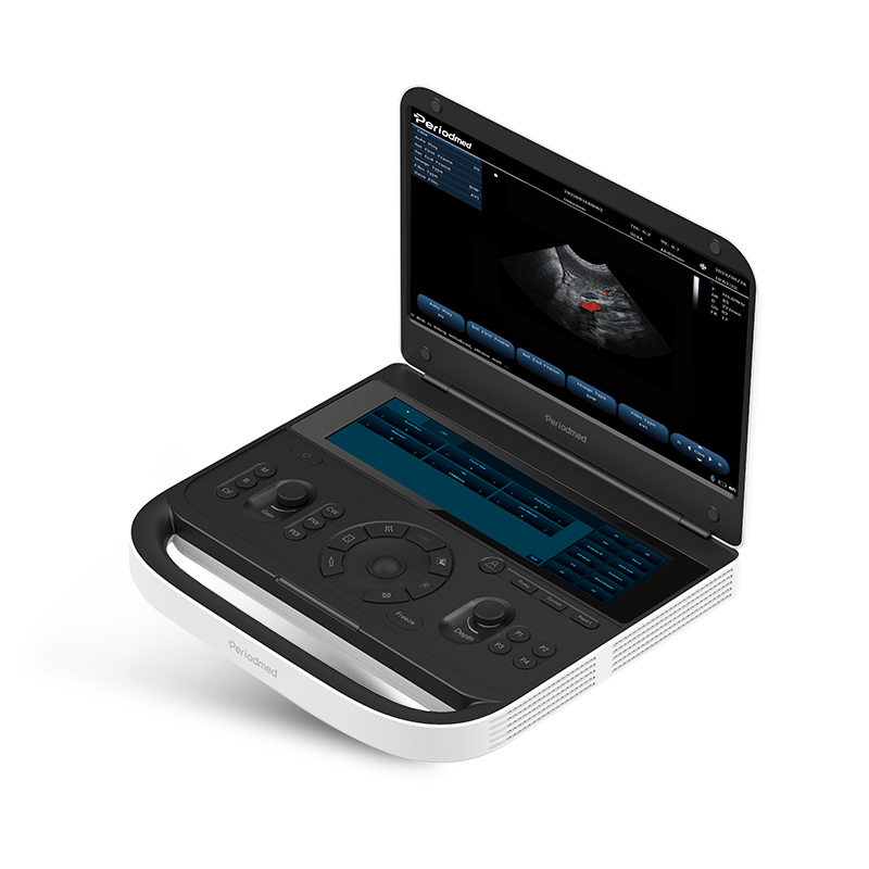
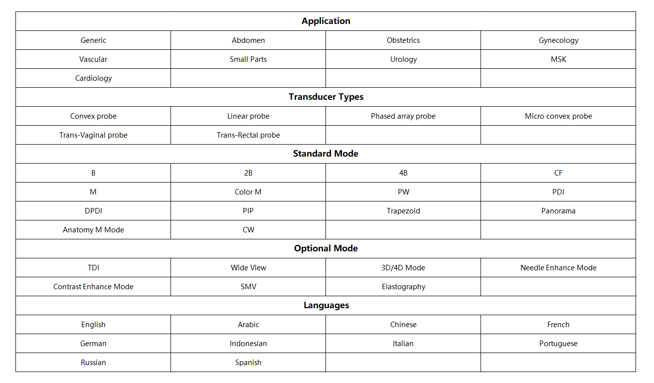
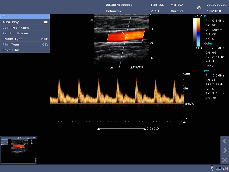
Features: 1. High-resolution imaging: using advanced ultrasound imaging technology, can provide high-resolution images to help doctors diagnose diseases accurately. 2. Many functions: has a variety of functions, including B/CF/M/PW/ CW/PDI/DPDI/TDI / 3 D / 4 D/wide scene imaging/puncture mode/contrast imaging mode, meet the needs of different departments. 3. Portable design: portable design, light weight, small size, convenient for doctors to move between different departments. 4. Friendly interface: with intuitive user interface and simple and easy to use operating system, so that doctors can get started quickly and make accurate diagnosis. 5. High performance sensors: Equipped with high performance sensors, able to provide clear images and accurate measurement results.
Key features
- Design highlights:
- 1. Ergonomic key operation: silicone interactive backlit keyboard, trackball
- 2. Double screen display: 12.6 inch full-size multi-function touch screen +15.6 inch anti-glare LED screen
- 3. The machine can connect two probes at the same time
- 4. Synchronization ability:
- B/CF, B/PDI or DPDI, B/PW, B/M, B+CF or PDI or DPDI/PW or M mode
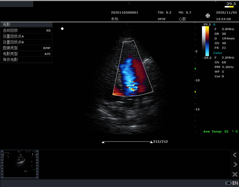
- 5. Echo enhancement technology Adaptive signal processing technology was used to analyze the echo signal of the undetermined region through a unique intelligent data sensing method, which improved the image resolution and uniformity, and easily obtained high- definition heart images
- 8.Composite imaging technology Allows the use of multiple scan space compartments, resulting in enhanced contrast and improved resolution visualization
- Probe Specifications:
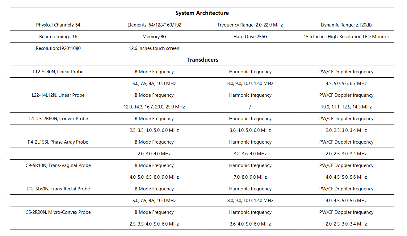
- 1. 2.0-10MHz V¬ariable frequency, frequency range 2.0-10MHz; 2. 5 kinds of frequencies of each probe, variable fundamental and harmonic frequency; 3. Abdomen: 2.5-6.0MHz; 4. Superficial:5.0-10MHz; 5. Cardiac:2.0-3.5MHz; 6. Puncture Guidance: probe puncture guide is optional, puncture line and Angle are adjustable; 7. Transvaginal: 5.0-9MHZ.
- 1. One-click optimization function
- Improved image quality based on automatic structure detection
- 2. rich echo beamforming technology
- The rich echo beam front allows the echo signal from adjacent beams that are traditionally ignored to be used to form a thinner, stronger imaging beam, providing better "out of focus" image resolution and deeper image penetration.
Applications
- Diagnostic ultrasound imaging in hospitals, clinics, emergency care, and mobile examination settings
Technical specifications
Design highlights:
1. Ergonomic key operation: silicone interactive backlit keyboard, trackball
2. Double screen display: 12.6 inch full-size multi-function touch screen +15.6 inch anti-glare LED screen
3. The machine can connect two probes at the same time
4. Synchronization ability:
B/CF, B/PDI or DPDI, B/PW, B/M, B+CF or PDI or DPDI/PW or M mode
5. Echo enhancement technology Adaptive signal processing technology was used to analyze the echo signal of the undetermined region through a unique intelligent data sensing method, which improved the image resolution and uniformity, and easily obtained high- definition heart images
6. Contrast imaging technology Using the second harmonic and nonlinear fundamental wave imaging, more accurate emission control to obtain excellent signal-to-noise ratio images. At the same time, it has microangiography imaging function and advanced angiographic quantitative analysis software, which provides more accurate judgment basis for clinical.
7. Elastography technology The latest elastography technology is used to improve the sensitivity of elastography, reduce the dependence on operation or manipulation, and show higher frame rate, better sensitivity, better stability and repeatability. 9, Harmonic imaging technology Using the second harmonics generated by the tissue boundary layer, THI significantly increases contrast resolution and improves image quality , especially for technically difficult subjects.
8.Composite imaging technology Allows the use of multiple scan space compartments, resulting in enhanced contrast and improved resolution visualization
Probe Specifications:
1. 2.0-10MHz V¬ariable frequency, frequency range 2.0-10MHz;
2. 5 kinds of frequencies of each probe, variable fundamental and harmonic frequency;
3. Abdomen: 2.5-6.0MHz;
4. Superficial:5.0-10MHz;
5. Cardiac:2.0-3.5MHz;
6. Puncture Guidance: probe puncture guide is optional, puncture line and Angle are adjustable;
7. Transvaginal: 5.0-9MHZ.
Optional Probes:
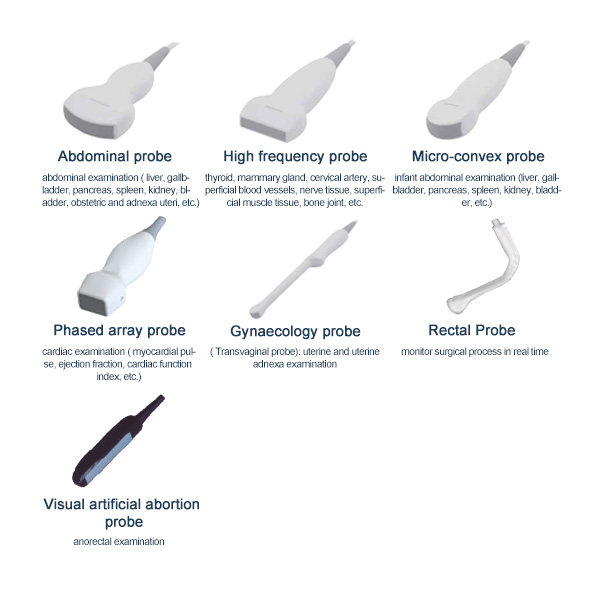
1. Abdominal probe: abdominal examination ( liver, gallbladder, pancreas, spleen, kidney, bladder, obstetric and adnexa uteri, etc.);
2. High frequency probe: thyroid, mammary gland, cervical artery, superficial blood vessels, nerve tissue, superficial muscle tissue, bone joint, etc.;
3.Micro-convex probe: infant abdominal examination (liver, gallbladder, pancreas, spleen, kidney, bladder, etc.);
4. Phased array probe: cardiac examination ( myocardial pulse, ejection fraction, cardiac function index, etc.);
5. Gynaecology probe (Transvaginal probe): uterine and uterine adnexa examination;
6. Visual artificial abortion probe: monitor surgical process in real time;
7. Rectal Probe: anorectal examination.
1. One-click optimization function
Improved image quality based on automatic structure detection
2. rich echo beamforming technology
The rich echo beam front allows the echo signal from adjacent beams that are traditionally ignored to be used to form a thinner, stronger imaging beam, providing better “out of focus” image resolution and deeper image penetration.
3. A maximum of 16 tasks for a transmission beam, resulting in excellent time resolution and higher frame rates.
4.Anatomical M-mode technology
Obtain accurate anatomical observations at any angle by freely placing sampling lines. Get better images with anatomical observation, up to three sampling lines
5.TDI: Tissue Doppler imaging allows you to quantitatively assess regional
myocardial motion and function, providing a complete TDI pattern for faster and more direct diagnosis.
Product video
An official product video is attached through the WooCommerce product-video metadata.
Ordering and configuration
Contact Scientific China to confirm the exact Yonker model, intended-use market, measurement parameters, included and optional accessories, compatible sensors or consumables, power supply, applicable certifications, minimum order quantity, current quotation, lead time, warranty, training, and international delivery terms.
Reviews (0)
Only logged in customers who have purchased this product may leave a review.


































 No products in the cart.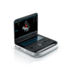
Reviews
There are no reviews yet.