


Live Cell Imaging System MCS31 for In-Incubator Cell Monitoring
Available in stock
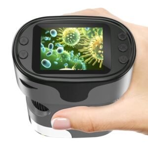
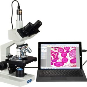
The Live Cell Imaging System MCS31 is an advanced in-incubator live cell imaging system designed for long-term cell monitoring, fluorescence imaging, and phase contrast observation. Compatible with culture flasks, dishes, and multi-well plates from 6-well to 384-well, it supports automated time-lapse recording, region scanning, image stitching, autofocus, and cell analysis. Ideal for cell culture, drug screening, and biological research, the MCS31 helps improve workflow efficiency while reducing contamination risk.
Category: Microscopes

Report Abuse
Scientific China
Store
Scientific China
StorePeople Also Viewed
-
 Cedrec FA1004B External Calibration Analytical Balance
$230.00
Cedrec FA1004B External Calibration Analytical Balance
$230.00
-
 Feline Blood Typing Kit for Cats | Rapid Veterinary Blood Type Test
$2.99
Feline Blood Typing Kit for Cats | Rapid Veterinary Blood Type Test
$2.99
-
 Touch Screen Constant-Temperature Incubator – BIOBASE CO₂ Incubator
-
 JLO-1090Si Stationary Oxygen Concentrator
-
 0.5ml Yellow Top Gel and Clot Activator Sst Blood Collection Microtubes
$0.02
0.5ml Yellow Top Gel and Clot Activator Sst Blood Collection Microtubes
$0.02
Questions & Answers
Loading...
Product Enquiry
Product Enquiry
Description
The Live Cell Imaging System MCS31 is a compact, incubator-compatible live cell imaging platform designed for long-term cell culture observation, fluorescence imaging, and phase contrast monitoring. Built for researchers who need stable, non-invasive imaging over time, the MCS31 supports a wide range of vessels including culture flasks, petri dishes, and 6-well to 384-well plates.
Ideal for cell biology, cancer research, drug screening, stem cell studies, microbiology, and comparative experiments, this system enables automated time-lapse imaging, region scanning, image stitching, and multi-well plate recording without repeatedly removing samples from the incubator. By reducing handling and exposure, it helps minimize contamination risk while improving consistency and experimental efficiency.
With support for brightfield phase contrast, customizable fluorescence channels, motorized objective switching, autofocus, and intelligent cell analysis functions, the MCS31 live cell imaging system offers a practical and efficient solution for modern laboratories seeking reliable in-incubator cell imaging and long-term live cell monitoring.
Specifications
| Item | Specifications |
|---|---|
| Brightfield Observation | Transmitted phase contrast illumination, working distance 55mm 50,000-hour lifespan, 625nm low-phototoxicity LED light source |
| Fluorescence Observation (Customizable Wavelengths) |
Blue (B): 472–495nm Green (G): 543–560nm Ultraviolet (UV): 393–416nm |
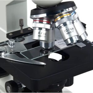
| Objective Lenses | Select any two from 4× / 10× / 20×; motorized switching |
| Stage | Motorized XY platform (115 × 77mm); repeat positioning accuracy ≤ 3μm |
| Z-axis | Motorized autofocus mechanism |
| Camera | High-speed, high-sensitivity camera; 5MP (500W), 2/3″ sensor, 40fps |
| Software | Remote control support: lighting, camera, image capture, video recording, time-lapse imaging |
| Analysis Functions | Cell counting, confluency analysis, scratch assay, transfection efficiency, viability analysis |
| Scanning Functions | Multi-well imaging: customizable XY array, multi-point capture, brightfield & fluorescence overlay Region scanning: rectangular area stitching, multi-channel overlay |
| Data Transmission / Power | USB data cable + DC power supply |
| Working Environment | 5–40°C, 20–95% RH |
| Dimensions | 252 × 377 × 208 mm |
Reviews (0)
Only logged in customers who have purchased this product may leave a review.
 No products in the cart.
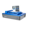
Reviews
There are no reviews yet.